. 2017 May:106:62-68.
doi: 10.1016/j.freeradbiomed.2017.02.020. Epub 2017 Feb 9.

Isolevuglandins as a gauge of lipid peroxidation in human tumors

Affiliations

Isolevuglandins as a gauge of lipid peroxidation in human tumors

H P Yan et al. Free Radic Biol Med. 2017 May.

Abstract

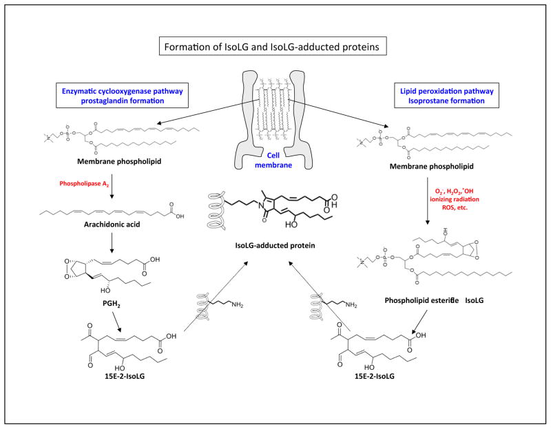
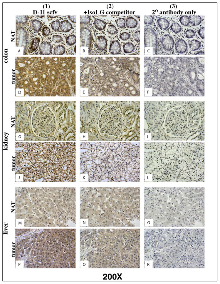
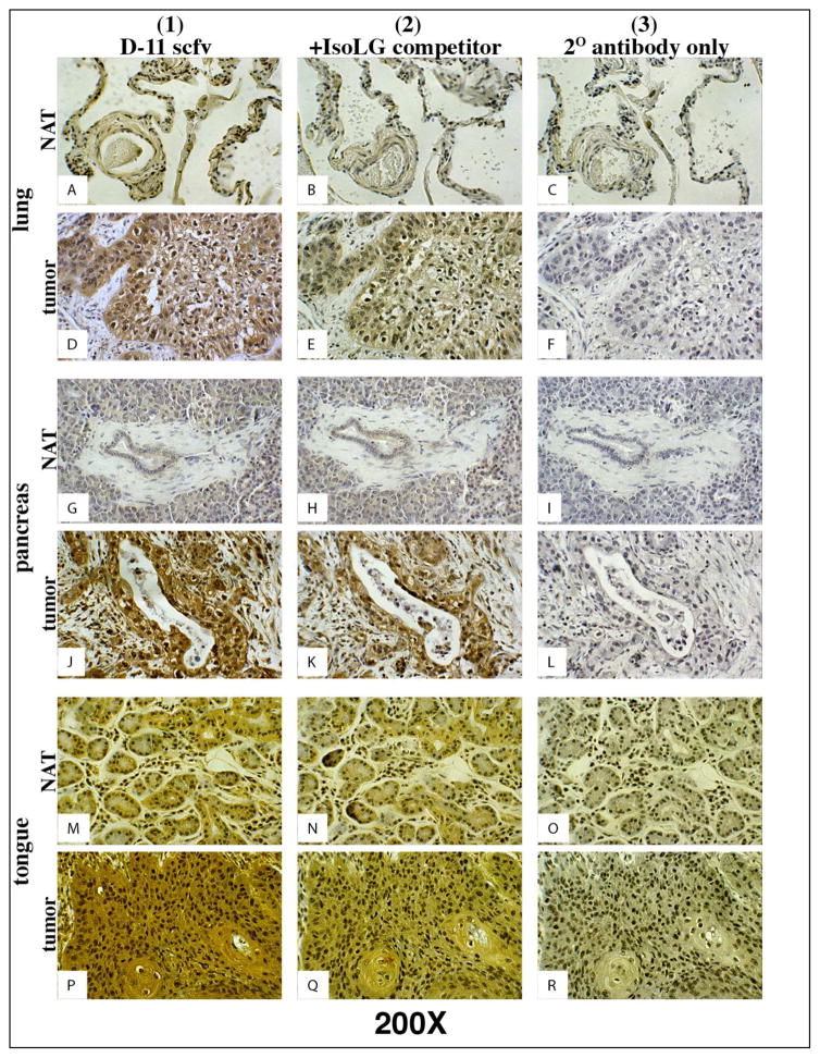
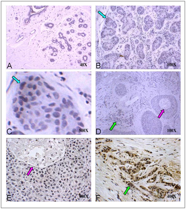
The cellular production of free radicals or reactive oxygen species (ROS) can lead to protein, lipid or DNA modifications and tumor formation. The cellular lipids undergo structural changes through the actions of enzymes (e.g. cyclooxygenases) or free radicals to form a class of compounds called Isolevuglandins (IsoLGs). The recruitment and continued exposure of tissue to ROS and IsoLGs causes increased cell proliferation, mutagenesis, loss of normal cell function and angiogenesis. The elevated concentration of ROS in cancerous tissues suggests that these mediators play an important role in cancer development. We hypothesized that tumors with elevated ROS levels would similarly possess an increased concentration of IsoLGs when compared with normal tissue. Using D11, an ScFv recombinant antibody specific for IsoLGs, we utilized immunohistochemistry to visualize the presence of IsoLG in human tumors compared to normal adjacent tissue (NAT) to the same tumor. We found that IsoLG concentrations were elevated in human breast, colon, kidney, liver, lung, pancreatic and tongue tumor cells when compared to NAT and believe that IsoLGs can be used as a gauge indicative of lipid peroxidation in tumors.

Keywords: Free radicals; Human tumors; Isolevuglandins; Lipid peroxidation.


Conflict of interest statement

Conflict of Interest Statement

The authors have no conflicts of interest.

Figures

Figure 1
Figure 1. Formation of isolevuglandin (IsoLG) and IsoLG protein adducts
Two pathways are used to form IsoLG protein adducts. The enzymatically derived prostaglandin pathway (left) and the free radical (lipid peroxidation) derived isoprostane pathway (right) and are depicted. (Left) Phospholipase A2 (PLA2) can cleave phospholipids to form arachidonic acid. Cyclooxygenase can oxidize arachidonic acid to form prostaglandin H2 that can subsequently undergo chemical rearrangement to form IsoLG (e.g. the 15E-2-IsoLG regioisomer). (Right) ROS stemming from mitochondria in a hypoxic tumor microenvironment can lead to lipid peroxidation. Oxidized lipids can undergo non-enzymatic chemical rearrangement to form IsoLG and IsoLG protein adducts. The chemically reactive IsoLG can then adduct to free amines (-NH2) on proteins or DNA (not depicted).
Figure 2
Figure 2. D11 scfv staining on serial colon, kidney and liver normal and tumor tissue
Column 1) Tissues stained with D11 scfv; Column 2) Negative control - Tissues stained with the D11 scfv pre-incubated with a competing concentration of IsoLG-adducted albumin; Column 3) Negative control - Tissues stained only with the Anti-E tag/HRP secondary (2o) antibody. Note: Tumor cells in column 1 stain (brown) more intensely than normal (NAT) cells adjacent to the tumor. Magnification for all images depicted is 200X.
Figure 3
Figure 3. D11 scfv staining on serial lung, pancreas and tongue normal and tumor tissue
Column 1) Tissues stained with D11 scfv; Column 2) Negative control - Tissues stained with the D11 scfv pre-incubated with a competing concentration of IsoLG-adducted albumin; Column 3) Negatvie control - Tissues stained only with the Anti-E tag/HRP secondary (2o) antibody. Note: Tumor cells in column 1 stain (brown) more intensely than normal (NAT) cells adjacent to the tumor. Magnification for all images depicted is 200X.
Figure 4
Figure 4. D11 scfv staining on normal, hyperplasia and malignant breast tissue
A) Normal breast tissue at low (40X) magnification; B) Breast hyperplasia (100X); C) Benign hyperplasia at high (1,000X) magnification (blue arrows in B and C point to the same site at different magnifications); D) In situ and invasive breast cancer at low (40X) magnification; E) In situ carcinoma at 400X magnification (purple arrows in D and E point to the same site at different magnifications). F) Invasive cancer at 400X magnification (green arrows in D and F point to the same site at different magnifications). Note: only the invasive carcinoma in Panel F shows strong D11 scfv (brown) staining.

References

    1. Rakoff-Nahoum S. Why cancer and inflammation? Yale J Biol Med. 2006;79:123–130. - PMC - PubMed
    1. Coussens LM, Werb Z. Inflammation and cancer. Nature. 2002;420:860–867. - PMC - PubMed
    1. Shacter E, Weitzman SA. Chronic inflammation and cancer. Oncology (Williston Park) 2002;16:217–226. 229. discussion 230–212. - PubMed
    1. Paravicini TM, Touyz RM. NADPH oxidases, reactive oxygen species, and hypertension: clinical implications and therapeutic possibilities. Diabetes Care. 2008;31(Suppl 2):S170–180. - PubMed
    1. Van der Paal J, Neyts EC, Verlackt CCW, Bogaerts A. Effect of lipid peroxidation on membrane permeability of cancer and normal cells subjected to oxidative stress. Chem Sci. 2016;7:489–498. - PMC - PubMed

Publication types

MeSH terms

LinkOut - more resources