. 2019 Apr;104(4):411-425.
doi: 10.1007/s00223-018-0502-5. Epub 2018 Dec 4.

Trauma-Induced Nanohydroxyapatite Deposition in Skeletal Muscle is Sufficient to Drive Heterotopic Ossification

Affiliations

Trauma-Induced Nanohydroxyapatite Deposition in Skeletal Muscle is Sufficient to Drive Heterotopic Ossification

Stephanie N Moore-Lotridge et al. Calcif Tissue Int. 2019 Apr.

Abstract

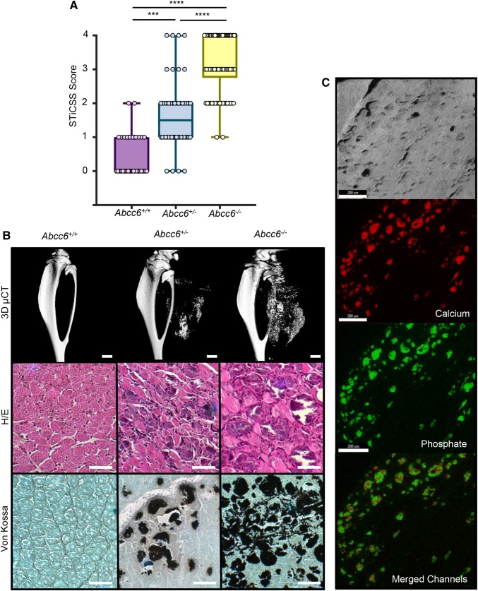
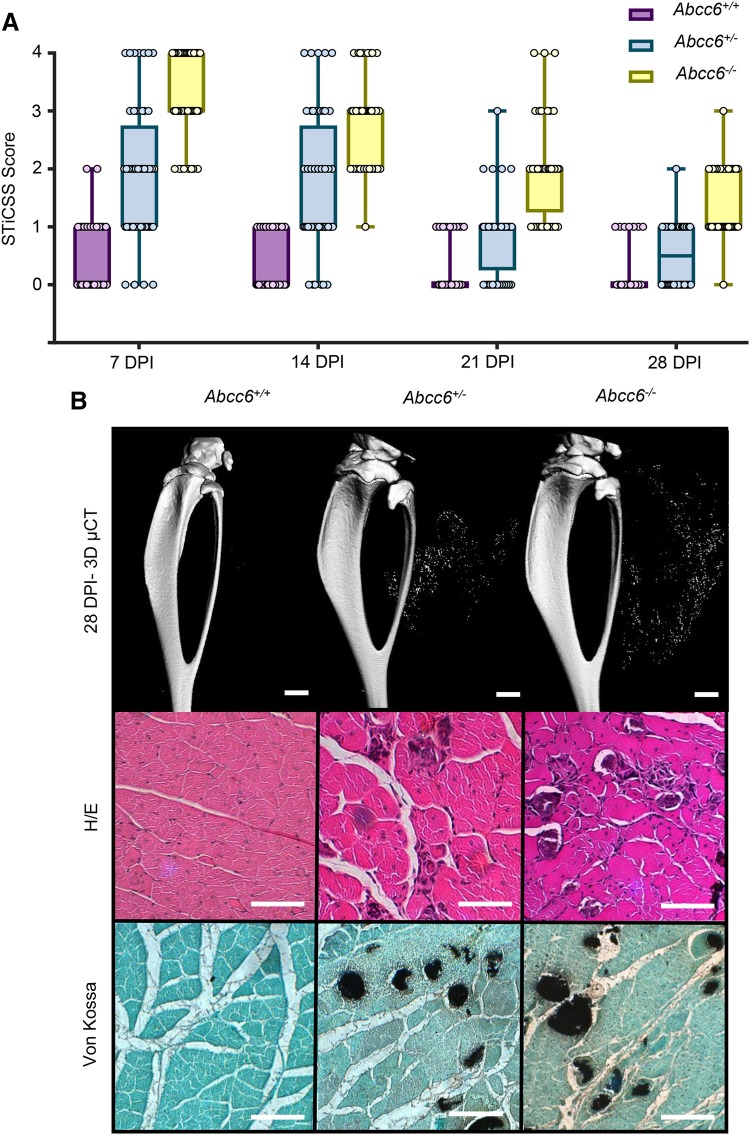
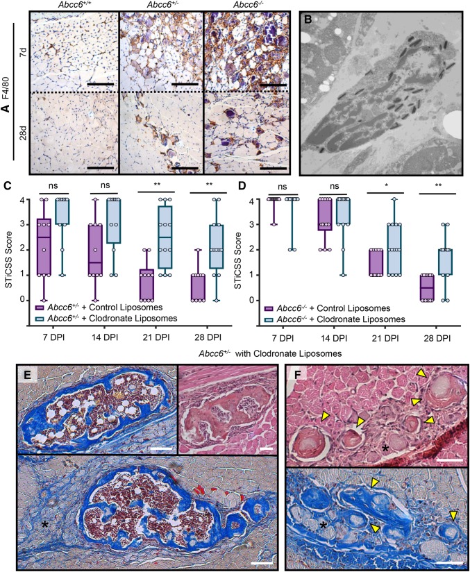
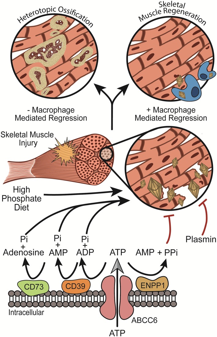
Heterotopic ossification (HO), or the pathologic formation of bone within soft tissues, is a significant complication following severe injuries as it impairs joint motion and function leading to loss of the ability to perform activities of daily living and pain. While soft tissue injury is a prerequisite of developing HO, the exact molecular pathology leading to trauma-induced HO remains unknown. Through prior investigations aimed at identifying the causative factors of HO, it has been suggested that additional predisposing factors that favor ossification within the injured soft tissues environment are required. Considering that chondrocytes and osteoblasts initiate physiologic bone formation by depositing nanohydroxyapatite crystal into their extracellular environment, we investigated the hypothesis that deposition of nanohydroxyapatite within damaged skeletal muscle is likewise sufficient to predispose skeletal muscle to HO. Using a murine model genetically predisposed to nanohydroxyapatite deposition (ABCC6-deficient mice), we observed that following a focal muscle injury, nanohydroxyapatite was robustly deposited in a gene-dependent manner, yet resolved via macrophage-mediated regression over 28 days post injury. However, if macrophage-mediated regression was inhibited, we observed persistent nanohydroxyapatite that was sufficient to drive the formation of HO in 4/5 mice examined. Together, these results revealed a new paradigm by suggesting the persistent nanohydroxyapatite, referred to clinically as dystrophic calcification, and HO may be stages of a pathologic continuum, and not discrete events. As such, if confirmed clinically, these findings support the use of early therapeutic interventions aimed at preventing nanohydroxyapatite as a strategy to evade HO formation.

Keywords: Abcc6; Dystrophic calcification; Heterotopic ossification; Nanohydroxyapatite; Skeletal muscle injury.


Conflict of interest statement

Conflict of interest

Jonathan G. Schoenecker is a member of the education advisory board at OrthoPediatrics and receives research funding from OrthoPediatrics and research support from IONIS Pharmaceuticals. Jonathan G. Schoenecker receives research support from PXE International. Stephanie N. Moore-Lotridge, Qiaoli Li, Breanne H. Y. Gibson, Joseph T. Martin, Gregory D. Hawley, Thomas H. Arnold, Masanori Saito, Sami Tannouri, Herbert S. Schwartz, Richard J. Gumina, Justin M.M. Cates, Jouni Uitto, and Jonathan G. Schoenecker have declared that no conflict of interest exists.

Human and Animal Rights and Informed Consent

All animal procedures were approved by the Vanderbilt University IACUC (M1600225). All procedures performed in studies involving animals were in accordance with the ethical standards of the institution at which the studies were conducted. Welfare-related assessments were carried out prior to and throughout all experiments by trained personnel at Vanderbilt University and Vanderbilt University Medical Center. This article does not contain any studies with human participants performed by any of the authors.

Figures

Fig. 1
Fig. 1
Loss of ABCC6 predisposes skeletal muscle to nanohydroxyapatite deposition at 7DPI. a WT (Abcc6+/+), heterozygous (Abcc6+/−), and homozygous (Abcc6−/−) mice were assessed for calcification at the site of skeletal muscle injury by radiographic analysis and subsequent STiCSS quantification at 7 DPI. See Table 2 for detailed analysis of the genotypes and N. ***p < 0.001; ****p < 0.0001. Statistical analysis between groups was performed using a non-parametric Mann–Whitney test. b Representative 3D µCT reconstructions and histological analysis of skeletal muscle calcification within the injured gastrocnemius and soleus muscles at 7 DPI. Scale bar represents 100 µm. n ≥ 3 mice per genotype. Positive Von Kossa staining, noted by the black deposits, indicates calcium deposition within the damaged skeletal muscle. c Energy dispersive X-ray (EDS) analysis of dystrophic calcification nodules within damaged ABCC6-deficient skeletal muscle at 14 DPI. Topographic mapping demonstrated marked co-localization of calcium and phosphate with an average calcium/phosphate atomic ratio of 1.67 ± 0.2, indicative of hydroxyapatite. Analysis was conducted following random sampling of 5 distinct spots per tissue section. Scale bar represents 200 µM, thereby indicating nanohydroxyapatite
Fig. 2
Fig. 2
Nanohydroxyapatite deposition in ABCC6-Deficient Mice is Degraded Over 28 DPI. Beginning at 7DPI, Abcc6+/+, Abcc6+/−, or Abcc6−/− animals were a assessed weekly by radiographic analysis through 28 DPI and quantified by the STiCCS, to reveal progressive resolution of nanohydroxyapatite from damaged skeletal muscle. b 3D µCT and histologic analysis at 28 DPI demonstrates reduced nanohydroxyapatite deposition compared to results seen in 7 DPI. H/E staining was utilized to assess sarcomere morphology and regeneration quantified in Table 2, and Von Kossa Staining was used to visualize calcification. Scale bar represents 100 µm. n ≥ 3 mice per genotype
Fig. 3
Fig. 3
Macrophage-mediated resolution of nanohydroxyapatite prevents maturation to HO. a Immunohistochemical stain for F4/80+ cells at 7 and 28 DPI in injure skeletal muscle from Abcc6+/+, Abcc6+/−, or Abcc6−/− mice. b Transition electron microscope image of a macrophage containing phagocytosed nanohydroxyapatite. Scale bar represents 500 nm. Image was obtained from a WT C57BL/6J following CTX injury at 3 days post injury when macrophage infiltration to damaged tissue is greatest [36]. c Longitudinal STiCSS analysis of Abcc6+/− treated with either control (PBS) or clodronate-filled liposomes beginning at the time of injury. N ≥ 5 mice per group. Nanohydroxyapatite was not observed in Abcc6+/+ mice treated with either control or clodronate-filled liposomes, N ≥ 4 mice per group. Data not shown. **p < 0.01; ***p < 0.001. d MSB histological analysis at 28 DPI of Abcc6+/− mice treated with control liposomes. e, f Histological analysis of Abcc6+/− mice treated with clodronate-containing liposomes indicating the presence of mature HO (E, MSB, and H/E) characterized by the presence of woven bone, blue staining (MSB) indicative of collagen deposition, and central hematopoiesis. Regions of persistent nanohydroxyapatite (black asterisks), and regions that appear to be nanohydroxyapatite maturing to ossified lesions (F-Yellow arrows, MSB, and H/E) were also observed. Scale bar represents 100 µm
Fig. 4
Fig. 4
The “Two Hit” mechanism of HO formation: taken together with previous studies [36], our results suggest that nanohydroxyapatite can deposit with in skeletal muscle following injury if one of the skeletal muscle protection mechanisms (i.e., ABCC6 or plasmin) are insufficient. Fortunately, the body possesses a secondary macrophage-mediated clean-up crew to regress nanohydroxyapatite from damaged tissues, thereby resolving the predisposing factors to HO formation. Together, these two lines of defense are critical for preventing nanohydroxyapatite deposition within damaged tissues and its subsequent maturation to HO. These findings suggest a new paradigm for HO formation in which HO can result from insufficient protection against nanohydroxyapatite with a failure of macrophage-mediated regression

References

    1. Ranganathan K, Loder S, Agarwal S, Wong VW, Forsberg J, Davis TA, Wang S, James AW, Levi B. Heterotopic ossification: basic-science principles and clinical correlates. J Bone Joint Surg Am. 2015;97(13):1101–1111. - PMC - PubMed
    1. Nauth A, Giles E, Potter BK, Nesti LJ, O’Brien FP, Bosse MJ, Anglen JO, Mehta S, Ahn J, Miclau T, Schemitsch EH. Heterotopic ossification in orthopaedic trauma. J Orthop Trauma. 2012;26(12):684–688. - PMC - PubMed
    1. Garland DE (1988) Clinical observations on fractures and heterotopic ossification in the spinal cord and traumatic brain injured populations. Clin Orthop Relat Res (233):86–101 - PubMed
    1. Giannoudis PV, Grotz MR, Papakostidis C, Dinopoulos H. Operative treatment of displaced fractures of the acetabulum. A meta-analysis. J Bone Joint Surg Br. 2005;87(1):2–9. - PubMed
    1. Dey D, Wheatley BM, Cholok D, Agarwal S, Yu PB, Levi B, Davis TA. The traumatic bone: trauma-induced heterotopic ossification. Transl Res. 2017;186:95–111. - PMC - PubMed

Publication types

Substances