. 2018 Dec:74:101-120.
doi: 10.1016/j.matbio.2018.07.004. Epub 2018 Jul 5.

A scar-like lesion is apparent in basement membrane after wound repair in vivo

Affiliations

A scar-like lesion is apparent in basement membrane after wound repair in vivo

William Ramos-Lewis et al. Matrix Biol. 2018 Dec.

Abstract

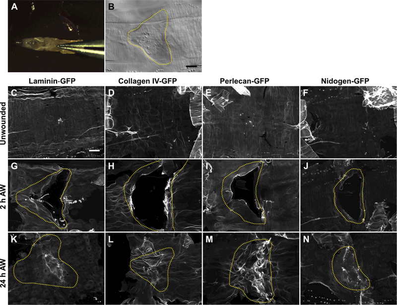
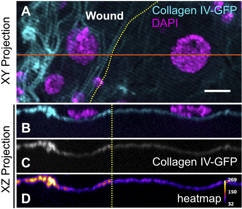
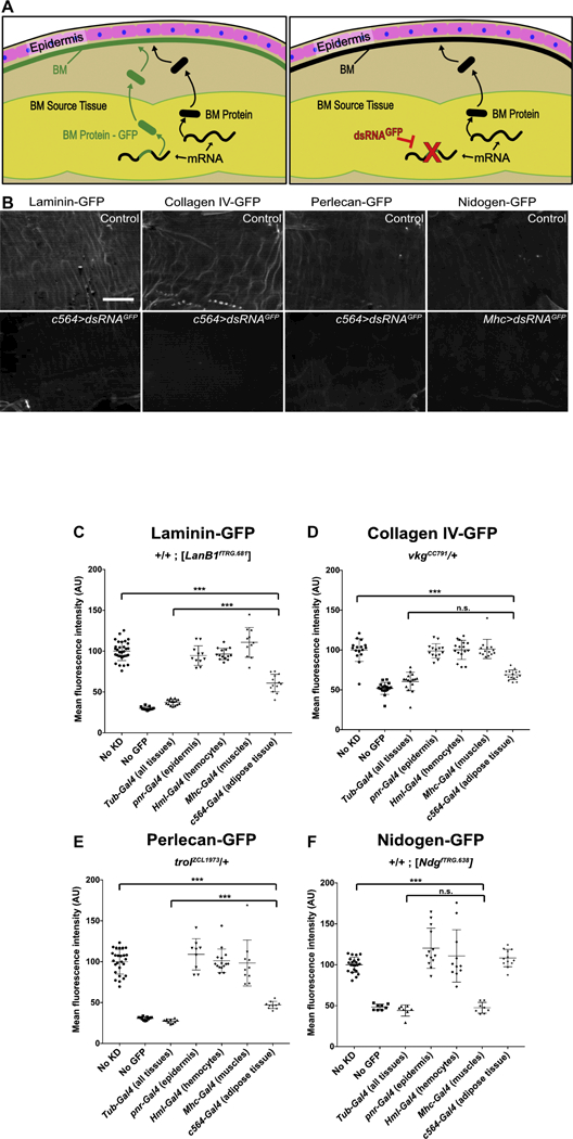
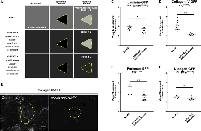
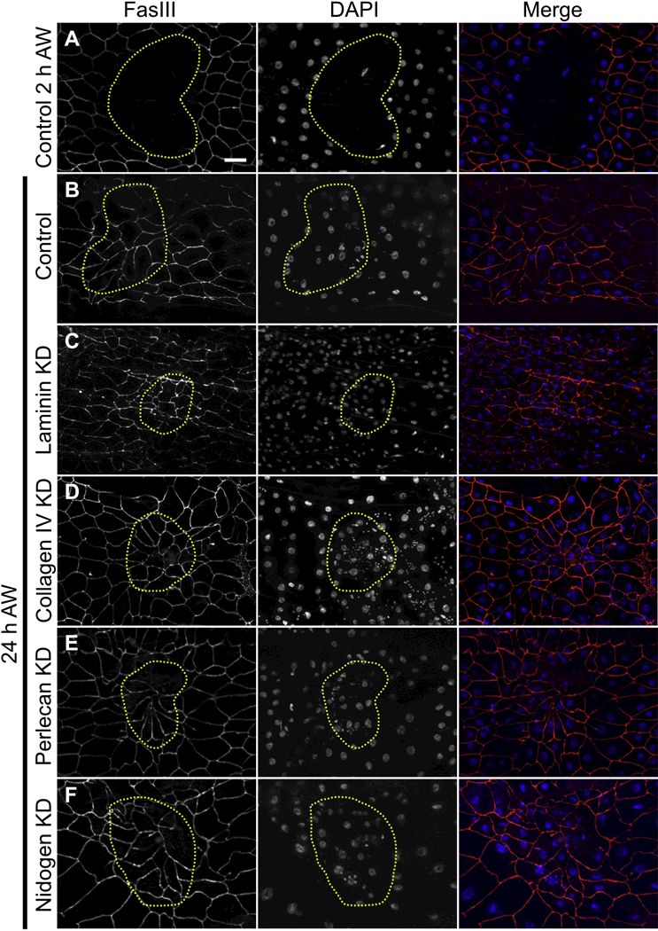
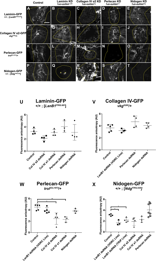
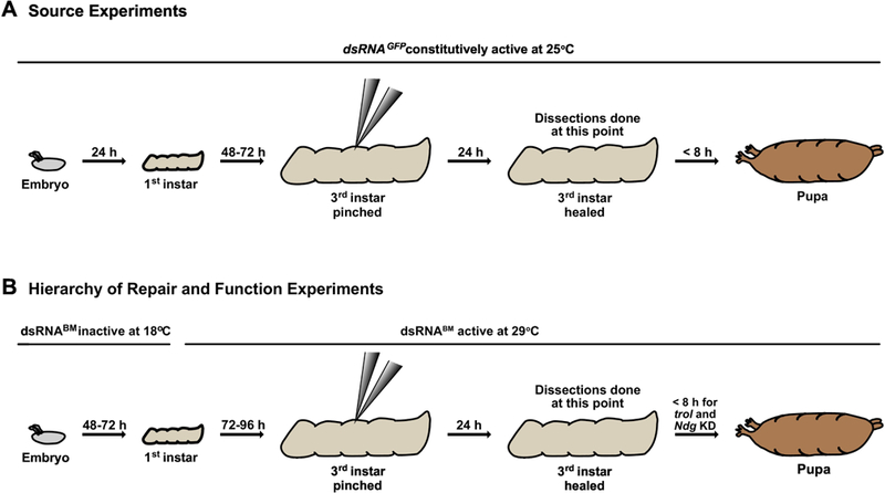
Basement membrane is a highly conserved sheet-like extracellular matrix in animals, underlying simple and complex epithelia, and wrapping around tissues like muscles and nerves. Like the tissues they support, basement membranes become damaged by environmental insults. Although it is clear that basement membranes are repaired after damage, virtually nothing is known about this process. For example, it is not known how repaired basement membranes compare to undamaged ones, whether basement membrane components are necessary for epithelial wound closure, or whether there is a hierarchy of assembly that repairing basement membranes follow, similar to the hierarchy of assembly of embryonic basement membranes. In this report, we address these questions using the basement membrane of the Drosophila larval epidermis as a model system. By analyzing the four main basement membrane proteins - laminin, collagen IV, perlecan, and nidogen - we find that although basement membranes are repaired within a day after mechanical damage in vivo, thickened and disorganized matrix scars are evident with all four protein components. The new matrix proteins that repair damaged basement membranes are provided by distant adipose and muscle tissues rather than by the local epithelium, the same distant tissues that provide matrix proteins for growth of unwounded epithelial basement membranes. To identify a hierarchy of repair, we tested the dependency of each of the basement membrane proteins on the others for incorporation after damage. For proper incorporation after damage, nidogen requires laminin, and perlecan requires collagen IV, but surprisingly collagen IV does not to depend on laminin. Thus, the rules of basement membrane repair are subtly different than those of de novo assembly.


Figures

Figure 1:
Figure 1:. The basement membrane is damaged by pinch wounds and forms a scar upon repair.
A) To damage the basement membrane, blunt forceps were used to pinch larvae on the dorsal epidermis between the hair stripes of segments A3 and A4. B) Pinch wounds do not break the outer cuticle, but they do leave an indentation visible with DIC, outlined with yellow dotted line. C-F) Undamaged epidermal basement membrane visualized with GFP-fusion constructs of each of the core basement membrane proteins. Images are representative of the no-knockdown controls quantified in Fig. 3. G-J) Damaged epidermal basement membrane after pinch-wounding. K-N) Within 24 h, the basement membrane was repaired, leaving behind a visible scar in the region of the healed wound. Images are representative of the no-knockdown controls quantified in Fig. 4. Dotted yellow lines indicate original wound borders based on cuticle indentation. Scale bar, 50 µm.
Figure 2:
Figure 2:. The basement membrane scar is thicker than unwounded basement membrane.
Yellow dotted lines indicate wound border. Orange solid line indicates location sampled for XZ projections. A) Basement membrane scar, evident on the left (wounded) side. B-D) Z-section shows increased thickness and fluorescence of collagen IV within the healed wound (N ≥ 3). Scale bar, 10µm.
Figure 3:
Figure 3:. In unwounded epidermis, basement membrane proteins come from other tissues.
A) Experiment overview: tissue expressing a basement membrane protein allele fused to GFP (BM-GFP) and an allele not fused with GFP will secrete both forms for incorporation into the basement membrane, resulting in fluorescent basement membrane. When dsRNAGFP targets GFP in the source tissue, only the basement membrane protein lacking GFP will be secreted, resulting in non-fluorescent basement membrane. B) Example images without (top) and with (bottom) dsRNAGFP expression. Scale bar, 50 µm. C-E) Laminin-GFP, collagen IV-GFP, or perlecan-GFP is lost from the epidermal BM when dsRNAGFP is expressed in adipose tissue. F) Nidogen-GFP is lost from the epidermal BM when dsRNAGFP is expressed in the muscles. *** indicates p ≤ 0.001.
Figure 4:
Figure 4:. The sources of basement membrane for repairing damage are the same as for growth.
A) Schematic of possible outcomes to test for a wound-specific source of basement membrane. In control unwounded epidermis, the basement membrane is fluorescent (depicted as medium gray color, top left) from the incorporation of BM-GFP. After repair, the basement membrane in the wound area is about 2-fold brighter than background (top right). If the tissue sources for growth (unwounded) and repair of basement membrane are different, then the permanent knockdown of GFP in the growth source tissue will cause the intact basement membrane (background) to become dim, but the repaired area is expected to remain bright, increasing the wound/background ratio. If the tissue source for growth (unwounded) and repair is the same, the permanent knockdown of GFP in the source tissue is expected to affect basement membrane repair in a similar manner, maintaining or reducing the wound/background ratio. B) Example of repaired epidermal basement membrane in the absence of dsRNAGFP (left) or in the presence of dsRNAGFP expressed in the fat body (right). C-F) Ratio of average fluorescence inside the wound over outside the wound with or without dsRNAGFP expression in the growth source tissue for epidermal basement membrane. Components appear to come from the same source tissues for growth and repair of basement membrane. * indicates p ≤ 0.05, ** indicates p ≤ 0.01. Scale bar, 50 µm.
Figure 5:
Figure 5:. Cells do not require any of the core basement membrane proteins to close wounds.
A) 2 h after injury, wounds were open. B-F) 24 h after injury, cells had closed the wound in (B) controls, or (C-F) when laminin, collagen IV, perlecan, or nidogen was knocked down days before wounding. Note that multinucleate syncytial cells were present after repair in control as well as knockdown wounds as previously reported [25]. Panels A-F are representative of 7, 25, 16, 15, 18, and 14 wounds examined, respectively. Scale bar, 50 µm.
Figure 6:
Figure 6:. Hierarchy of basement membrane assembly during repair.
A-E) Laminin assembled into repaired basement membrane independent of any other basement membrane proteins. F-J) Collagen IV assembled into repaired basement membrane independent of any other basement membrane proteins. K-O) Although perlecan assembled into repaired basement membrane independent of any other basement membrane proteins, its assembly into the scar required collagen IV (M). P-T) Nidogen required laminin (Q) but not collagen IV (R) or perlecan (S) to assemble into repaired basement membrane. In collagen knock-down wounds (C,H,M,R), scars appear to extend outside the wound area, see text. In panel Q, the bright dots at the wound center (marked by yellow stars) are autofluorescent melanization, see Experimental Procedures. U-X) Quantification of fluorescence anisotropy in repaired basement membranes. * indicates p ≤ 0.05, ** indicates p ≤ 0.01. Unless otherwise indicated, no significant difference was observed. Scale bar, 50 µm.
Figure 7:
Figure 7:. The requirement for laminin is different between basement membrane de novo assembly and repair.
A) Expression of dsRNA against LanB1 knocked down LanB1-GFP levels in stage 16– 17 embryos, as determined by GFP fluorescence. B) Quantification of GFP fluorescence in embryos showed dsRNA against LanB1 reduced LanB1-GFP levels by 68%. C) For comparison, expression of the same dsRNA against LanB1 in larvae depleted LanB1-GFP by 71% as measured by western blot (see also Fig. S3). D) Expression of dsRNA against LanB1 was sufficient to disrupt collagen IV deposition into the basement membrane of ventral nerve cord channels in stage 16–17 embryos, as visible in controls (yellow arrows) (N ≥ 3). Scale bar, 50 µm.
Figure 8:
Figure 8:. Timeline of experimental protocols for gene knockdown, showing larval development, dsRNA expression, and wounding.
A) For all basement membrane source experiments, animals were maintained at 25°C. Larvae developed to early 3rd instars, were pinched, and usually recovered 24 h before dissecting. For experiments where no wound was inflicted, larvae developed to late 3rd instar prior to dissecting. B) For basement membrane hierarchy of repair and function experiments in which LanB1, vkg, or trol was knocked down, embryos were laid and allowed to develop to 1st instar larvae at 18°C, with dsRNA not expressed. During 1st instar, bottles were shifted to 29°C to promote dsRNA expression, and larvae developed to early 3rd instar prior to wounding. After wounding, larvae recovered for 24 h at 29°C prior to dissecting. For Ndg KD experiments, bottles were maintained at 29°C for the entire experiment (not shown). Only control, trol KD and Ndg KD larvae were capable of pupariating. dsRNABM denotes dsRNA against vkg, trol, or LanB1.

References

    1. Fidler AL, Darris CE, Chetyrkin SV, Pedchenko VK, Boudko SP, Brown KL, et al. , Collagen IV and basement membrane at the evolutionary dawn of metazoan tissues, Elife 6 (2017) 1–24. . - DOI - PMC - PubMed
    1. Pozzi A, Yurchenco PD, Iozzo RV, The nature and biology of basement membranes, Matrix Biology 57–58 (2017) 1–11. . - DOI - PMC - PubMed
    1. Wang X, Harris RE, Bayston LJ, Ashe HL, Type IV collagens regulate BMP signalling in Drosophila, Nature 455 (2008) 72–77. . - DOI - PubMed
    1. Ma M, Cao X, Dai J, Pastor-Pareja JC, Basement Membrane Manipulation in Drosophila Wing Discs Affects Dpp Retention but Not Growth Mechanoregulation, Developmental Cell 42 (2017) 97–106.e4. . - DOI - PubMed
    1. Skeath JB, Wilson BA, Romero SE, Snee MJ, Zhu Y, Lacin H, The extracellular metalloprotease AdamTS-A anchors neural lineages in place within and preserves the architecture of the central nervous system, Development 144 (2017) 3102–3113. . - DOI - PMC - PubMed

Publication types

MeSH terms