. 2019 Apr 18;26(4):502-511.e3.
doi: 10.1016/j.chembiol.2018.12.003. Epub 2019 Jan 31.

Selection of DNA Cleavage Sites by Topoisomerase II Results from Enzyme-Induced Flexibility of DNA

Affiliations

Selection of DNA Cleavage Sites by Topoisomerase II Results from Enzyme-Induced Flexibility of DNA

Yunsu Jang et al. Cell Chem Biol. .

Abstract

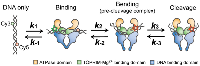
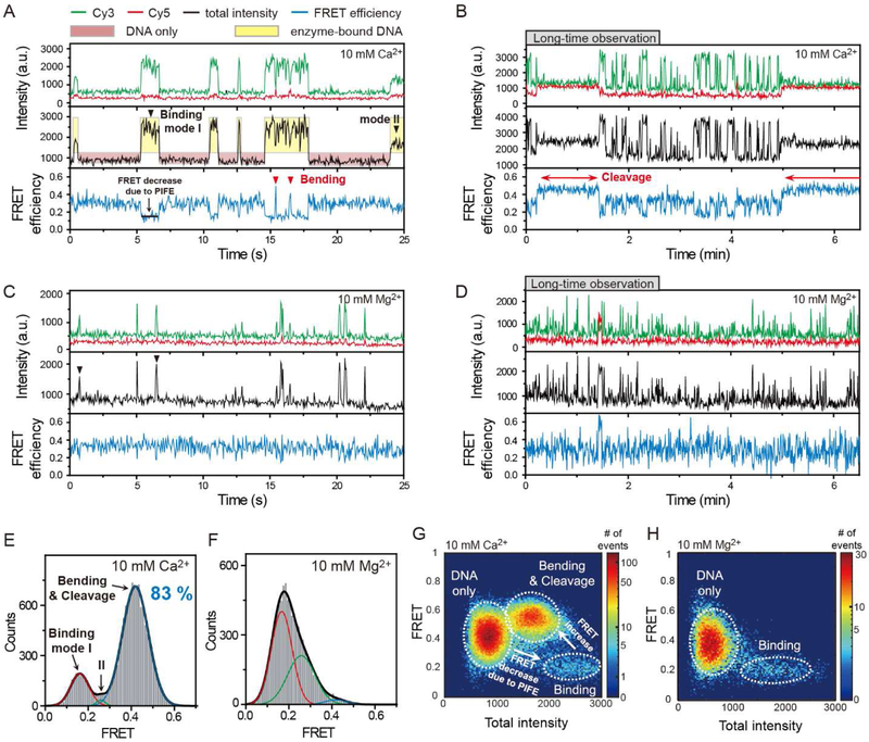
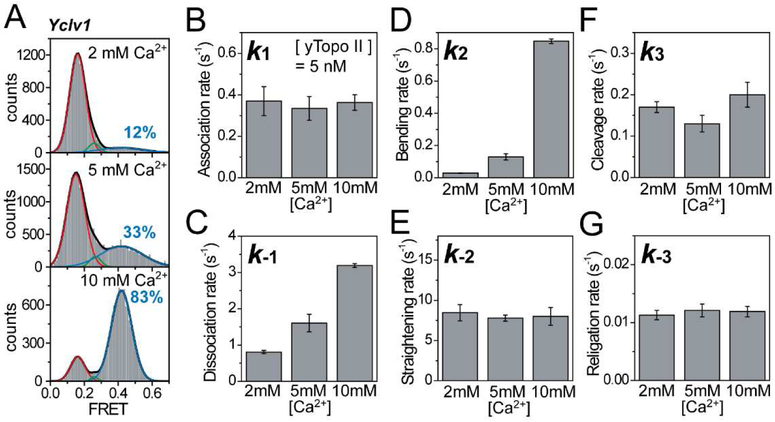
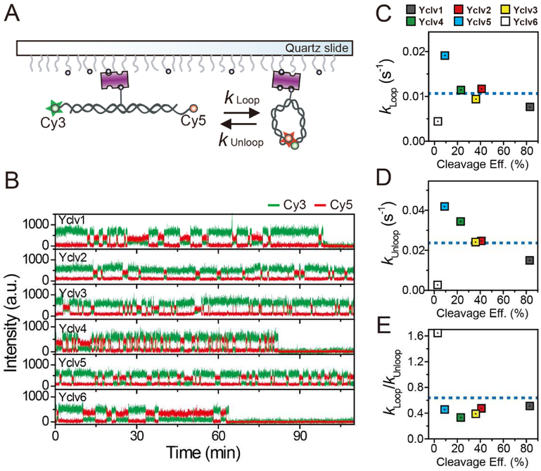
Topoisomerase II cleaves DNA at preferred sequences with different efficiencies; however, the mechanism of cleavage site selection is not known. Here we used single-molecule fluorescence assays that monitor several critical steps of DNA-topoisomerase II interactions, including binding/dissociation, bending/straightening, and cleavage/religation, and reveal that the cleavage site is selected mainly during the bending step. Furthermore, despite the sensitivity of the bending rate to the DNA sequence, it is not an intrinsic property of the DNA itself. Rather, it is determined by protein-DNA interactions.

Keywords: DNA bending; DNA cleavage; DNA flexibility; G-segment selection; sequence preference; single-molecule FRET; topoisomerase II; two-metal-ion mechanism.


Figures

Figure 1.
Figure 1.
Kinetic Scheme for the Topoisomerase II-mediated DNA Cleavage/Religation Reaction
Figure 2.
Figure 2.. Experimental Design for Observing DNA-topoisomerase II Interactions
(A) DNA construct (Yclv1) used for the experiments. The labeling sites for conjugating dyes, Cy3 and Cy5, are indicated. (B) Schematic diagram of single-molecule fluorescence resonance energy transfer (FRET) assay.
Figure 3.
Figure 3.. Real-time Observation of Topoisomerase II-mediated DNA Cleavage/Religation Reaction
(A,C) Representative time traces showing association/dissociation (black arrowheads) and bending/straightening events (red arrowheads) in the presence of 5 nM yTopoII and 10 mM Ca2+ (A) or Mg2+ (C) ions for Yclv1 DNA construct. In these measurements, a time resolution of 50 ms was used for observing transient interactions. Black arrowheads indicate transient binding of the enzyme to a DNA. Red arrowheads indicate the short-lasting bending events. Cy3 fluorescence, Cy5 fluorescence, and corresponding FRET efficiency are shown in green, red, and blue, respectively. Here, the total intensity is the sum of intensities of Cy3 and Cy5 and FRET efficiency (hereafter FRET) is the ratio of Cy5 intensity to the total intensity. To clearly visualize the enzyme binding events, red and yellow boxes are added as a visual guide. The same color convention is used throughout the paper. (B,D) Representative time traces showing DNA cleavage/religation (red arrows) events in the presence of 5 nM yTopoII and 10 mM Ca2+ (B) or Mg2+ (D) ions. In (B), experiment was performed under the same reaction condition as in (A) except for the time resolution. In (D), the experimental condition was same as in (C) except for the time resolution. For long-time observation, a time resolution of 500 ms was used to reduce the photobleaching of the fluorophores. The long-lived cleavage events are indicated by red arrows. (E,F) FRET histograms of Yclv1 DNA duplexes with bound enzyme in the presence of 5 nM yTopoII and 10 mM Ca2+ (E) or Mg2+ (F) ions. The cleavage population of Yclv1 (blue line, 83%) in the presence of Ca2+ ions was obtained by fitting the FRET histogram to the sum of three Gaussian functions. (G,H) Relative distribution of total intensity and FRET efficiency of Yclv1 DNA duplexes in the presence of 5 nM yTopoII and 10 mM Ca2+ (G) or Mg2+ (H) ions. The colors represent the relative frequency of the events. White dashed lines were added for a clear visualization of the three species, including unbound DNA only, enzyme binding, and DNA bending/cleavage.
Figure 4.
Figure 4.. Kinetic Pathway of Divalent Ions during the DNA Cleavage/Religation Reaction
(A) Fluorescence resonance energy transfer (FRET) histograms of DNA duplexes with bound enzyme at varying Ca2+ ion concentrations. Cleavage populations at varying Ca2+ ion concentrations were obtained by fitting the FRET histograms to the sum of three Gaussian functions. (B–G) Comparison of association (B), dissociation (C), bending (D), straightening (E), cleavage (F), and religation rates (G) at varying Ca2+ ion concentrations. Error bars represent standard deviations obtained from three independent experiments.
Figure 5.
Figure 5.. DNA Bending as a Main Determinant of the DNA Cleavage Efficiency
(A) Fluorescence resonance energy transfer (FRET) histograms of six DNA duplexes with bound enzyme in the presence of 10 mM Ca2+ ions. Cleavage populations of six DNA duplexes were obtained by fitting the FRET histograms to the sum of three Gaussian functions. (B–G) Comparison of association (B), dissociation (C), bending (D), straightening (E), cleavage (F), and religation rates (G) of DNA duplexes with different DNA base compositions. Error bars indicate standard deviations obtained from three independent experiments. To clearly visualize the corre-lation between each rate constant and the cleavage efficiency, linear fit lines (red line) and average values (blue dashed line) of the entire data set for each rate constant are added.
Figure 6.
Figure 6.. Intrinsic Bendability of the DNA Sequences
(A) Schematic diagram of the single-molecule cyclization assay. (B) Representative time traces of fluorescence intensity for six DNA duplexes with different DNA base compositions. All cyclization assays are conducted with 40 mM KCl and 1 mM MgCl2. For long-time observations, the excitation laser power was reduced as much as possible in spite of the low signal-to-noise ratio (~ 4) and a time resolution of 0.5 s was used to reduce the photobleaching of fluorophores. In this measurement, a theoretical limit of observation time was estimated to 12~120 minutes. The detailed estimation is provided in STAR Methods. (C–E) Comparison of looping rates (C), unlooping rates (D), and the ratio of the looping to unlooping rates (E) of DNA duplexes with different DNA base compositions. To clearly visualize the correlation between each rate constant and the cleavage efficiency, average values (blue dashed line) of the entire data set for each rate constant are added.

References

    1. Bax BD, Chan PF, Eggleston DS, Fosberry A, Gentry DR, Gorrec F, Giordano I, Hann MM, Hennessy A, Hibbs M, et al. (2010). Type IIA topoisomerase inhibition by a new class of antibacterial agents. Nature 466, 935–940. - PubMed
    1. Berger JM, Gamblin SJ, Harrison SC, and Wang JC (1996). Structure and mechanism of DNA topoisomerase II. Nature 379, 225–232. - PubMed
    1. Boedicker JQ, Garcia HG, Johnson S, and Phillips R (2013). DNA sequence-dependent mechanics and protein-assisted bending in repressor-mediated loop formation. Phys Biol 10, 066005. - PMC - PubMed
    1. Bromberg KD, Hendricks C, Burgin AB, and Osheroff N (2002). Human topoisomerase IIα possesses an intrinsic nucleic acid specificity for DNA ligation. Use of 5' covalently activated oligonucleotide substrates to study enzyme mechanism. J Biol Chem 277, 31201–31206. - PubMed
    1. Bromberg KD, and Osheroff N (2001). DNA cleavage and religation by human topoisomerase IIα at high temperature. Biochemistry 40, 8410–8418. - PubMed

Publication types

MeSH terms

Substances

LinkOut - more resources